Your Cells Are Batteries: The Electrical Nature of Life
Electricity in your body isn't just about neurons firing. Every cell maintains a voltage, and the body's bioelectric fields do things neuroscience is only beginning to map.

Your Cells Are Batteries: The Electrical Nature of Life
Series: Bioelectric Code | Part: 1 of 7 Primary Tag: FRONTIER SCIENCE Keywords: bioelectricity, membrane potential, ion pumps, cellular electricity, Vmem
The Basic Physics
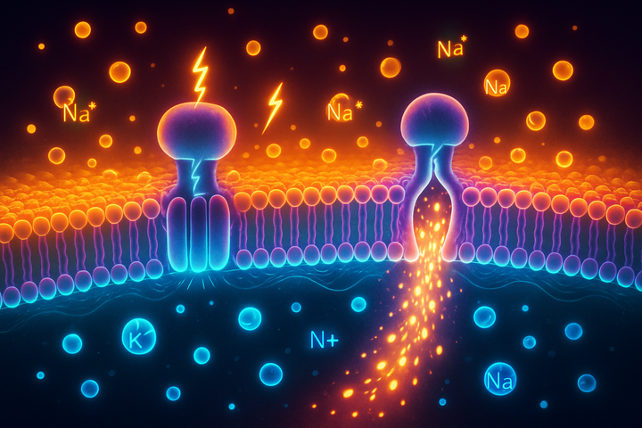
The electrical charge across a cell membrane arises from ion gradients—differences in the concentration of charged particles (ions) on either side of the membrane. The key players:
- Sodium (Na+): High concentration outside the cell, low inside
- Potassium (K+): High concentration inside the cell, low outside
- Chloride (Cl-): High concentration outside, low inside
- Calcium (Ca2+): Much higher outside, very low inside (calcium is tightly regulated)
The cell membrane is a lipid bilayer—essentially a wall of fat that ions can't freely cross. Embedded in this membrane are proteins that control ion movement: Ion pumps actively move ions against their concentration gradients, using ATP for energy. The most famous is the sodium-potassium pump (Na+/K+ ATPase), which pumps 3 sodium ions out for every 2 potassium ions pumped in. This pump runs constantly, consuming about 25% of your body's ATP, just to maintain ion gradients. Ion channels are gated pores that allow specific ions to flow down their concentration gradients when open. Channels can be gated by voltage, by chemical ligands, or by mechanical force. The combination of pumps (creating gradients) and channels (allowing controlled flow) produces the resting membrane potential: the voltage difference between inside and outside of the cell when it's not actively signaling.
Measuring Cellular Voltage
The resting membrane potential (often abbreviated Vmem or Vm) is typically negative—the inside of the cell is negative relative to the outside. Typical values:
- Neurons: -70 mV
- Muscle cells: -85 to -90 mV
- Epithelial cells: -30 to -60 mV
- Proliferating cells: -20 to -30 mV
- Stem cells: -10 to -30 mV
- Cancer cells: often depolarized (-10 to -25 mV)
That's millivolts—thousandths of a volt. Small in absolute terms, but cells are small. The electric field across a cell membrane is actually enormous—on the order of 10 million volts per meter. If you scaled it up, it would rival the electric field in a lightning bolt. We measure Vmem using patch-clamp electrophysiology (inserting a tiny glass electrode into cells) or voltage-sensitive fluorescent dyes that change brightness depending on local voltage. The key insight: Vmem varies by cell type and cell state. It's not fixed; it's regulated. And its value has consequences.
Voltage and Cell Fate
Here's where it gets interesting. The membrane potential isn't just a readout—it's a control variable. Proliferation vs. quiescence: Rapidly dividing cells (stem cells, cancer cells) tend to be depolarized (less negative Vmem). Quiescent, differentiated cells tend to be hyperpolarized (more negative Vmem). This isn't just correlation—artificially hyperpolarizing cells can push them toward differentiation; depolarizing can promote proliferation. Differentiation: Stem cells deciding their fate show characteristic voltage changes. Neural stem cells hyperpolarize as they become neurons. Muscle progenitors show voltage shifts before becoming mature muscle. The electrical state is part of the decision-making process. Migration: Cells migrating during wound healing orient themselves relative to local electrical fields (electrotaxis). Wounds generate voltage gradients; cells move toward the cathode (negative end). This isn't mystical—it's cells following bioelectric cues. Apoptosis: The cascade of events in programmed cell death includes membrane depolarization. The voltage change is part of how cells signal that they're dying. Voltage is information. The cell's electrical state tells it something about itself, its neighbors, and its context. And cells act on that information.
Beyond Single Cells: Tissue-Level Patterns
The bioelectric revolution really takes off when you consider tissues rather than individual cells. Cells in tissues are connected—by gap junctions, by electrical synapses, by shared extracellular space. Ion flow in one cell can affect voltage in neighboring cells. Tissues develop patterns of voltage—some regions more depolarized, others more hyperpolarized. These bioelectric patterns carry information about tissue organization. They're a distributed memory system, encoding anatomical information in a form that's independent of (though interacting with) genetic information. Michael Levin and colleagues have demonstrated this in remarkable experiments: Planarian head-tail polarity: Flatworms regenerate according to bioelectric patterns. Change the voltage pattern, and you can make a worm grow two heads, or no head, or heads at both ends. The voltage pattern encodes where the head should be. Eye induction: In frog embryos, certain ion channel manipulations can induce functional eyes to grow in aberrant locations—on the gut, on the tail. The bioelectric signature that says "make an eye here" can be artificially moved. Cancer normalization: Tumors have disrupted bioelectric properties. In some experiments, restoring normal voltage patterns can suppress tumor growth or normalize tumor cells toward normal behavior. The bioelectric layer is a coordination mechanism—a way for cells to know their position, their identity, and what they should become.
The Hardware: Ion Channels and Gap Junctions
The molecular machinery that creates bioelectric patterns includes: Voltage-gated channels: Ion channels that open or close depending on the local voltage. These create feedback loops—the voltage affects the channels which affect the ion flow which affects the voltage. Such circuits can have memory, oscillation, and other computational properties. Ligand-gated channels: Channels that open in response to specific molecules (neurotransmitters, signaling molecules). This links chemical signaling to electrical signaling. Gap junctions: Protein channels that directly connect the cytoplasm of adjacent cells. Ions (and small molecules) flow directly between cells through gap junctions, electrically coupling them. Gap junction patterns determine which cells share electrical signals. Transporters and pumps: The active machinery that establishes gradients. These require energy but allow cells to set their electrical state independently of what neighbors are doing. The same molecular toolkit used in the nervous system—ion channels, gap junctions, voltage-gated conductances—operates in all tissues. The nervous system is a specialized elaboration of a more general bioelectric communication system.
Why Did Biology Ignore This?
If bioelectricity is so important, why was it overlooked? Neuroscience captured the field. The study of bioelectricity became identified with the study of neurons. Action potentials, synaptic transmission, brain function—these were exciting and obviously electrical. The fact that all cells have electrical properties was known but treated as uninteresting. Molecular biology dominated. The central dogma—DNA → RNA → protein—focused attention on the genome as the information layer. Bioelectric information didn't fit the paradigm. It wasn't encoded in sequence; it wasn't heritable in the conventional sense (though its patterns are reconstructed each generation). Measurement challenges. Measuring voltage at the single-cell level requires specialized equipment. Measuring voltage patterns across tissues is harder still. The tools for bioelectric research were less accessible than those for molecular biology. It seemed like epiphenomenon. Even researchers who noticed non-neural bioelectricity often assumed it was a byproduct of cellular metabolism, not a causally important signal. Michael Levin, who has led much of the modern bioelectric revival, notes that the history of this field goes back to the 1940s-50s with researchers like Burr, Becker, and others. Their work was marginalized, then largely forgotten. The renaissance began in the 2000s with better tools and new experimental approaches.
The Bioelectric-Genetic Interface
Bioelectric signals don't replace genetic information—they work with it. Voltage affects gene expression. Ion channel activity can trigger signaling cascades that ultimately alter transcription. Depolarization can activate voltage-sensitive phosphatases, affect calcium entry (and thus calcium-dependent transcription factors), and modulate chromatin state. Genes encode bioelectric machinery. The ion channels, pumps, and gap junctions that create bioelectric patterns are genetically encoded. Mutations in these genes cause developmental abnormalities. Bioelectric patterns are epigenetic. Like chromatin modifications, bioelectric states carry information that isn't in the DNA sequence but influences how genes are used. They're a type of positional information—telling cells where they are and what they should become. The patterns self-organize. Unlike direct genetic control (gene A activates gene B), bioelectric patterns emerge from the interactions of many cells and channels. They're a distributed, emergent property. This gives them properties—robustness, regenerative capacity—that purely genetic control lacks. The genome is a parts list. Bioelectricity is part of the assembly instructions.
Implications
If every cell is electrical, and if bioelectric patterns coordinate development and regeneration, then: Disease may have bioelectric signatures. Cancer, birth defects, wound healing failures—these might have characteristic bioelectric abnormalities that could be diagnostic or therapeutic targets. Regeneration might be electrically triggered. Animals that regenerate (salamanders, planaria) have bioelectric patterns that enable regeneration. Could manipulating voltage patterns induce regeneration in animals (humans) that normally can't? Development might be bioelectrically controlled. Understanding the bioelectric code could allow reprogramming of developmental outcomes—correcting defects, or even building new tissues. Computing beyond neurons. If all cells compute with voltage, the body is a much richer computational substrate than we thought. The brain may be a specialized module in a body-wide bioelectric processing system. These are the topics we'll explore in this series.
Further Reading
- Levin, M. (2014). "Molecular bioelectricity: how endogenous voltage potentials control cell behavior and instruct pattern regulation in vivo." Molecular Biology of the Cell.
- Levin, M. (2021). "Bioelectric signaling: Reprogrammable circuits underlying embryogenesis, regeneration, and cancer." Cell.
- McLaughlin, K.A. & Bhavsar, R. (2021). "Microfluidic bioelectricity: Modeling the bioelectric microenvironment of cells." Annual Review of Biomedical Engineering.
- Becker, R.O. & Selden, G. (1985). The Body Electric. William Morrow.
This is Part 1 of the Bioelectric Code series, exploring your body's electrical nature. Next: "The Voltage of Life: Membrane Potential."


Comments ()