Cancer as Bioelectric Breakdown
Genetic mutations don't tell the whole story of cancer. A growing body of evidence points to bioelectric breakdown — changes in membrane voltage that corrupt a cell's identity and send it rogue.
Cancer as Bioelectric Breakdown
Series: Bioelectric Code | Part: 6 of 7 Primary Tag: FRONTIER SCIENCE Keywords: cancer, bioelectricity, depolarization, tumor microenvironment, ion channels, oncology
The Bioelectric Signature of Cancer
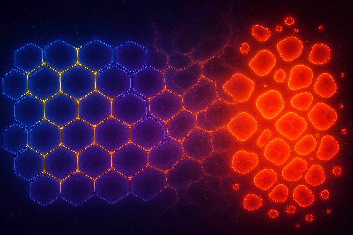
Across tissue types and cancer types, a consistent pattern emerges: cancer cells are depolarized. Normal cells maintain a negative resting membrane potential—typically between -40 and -90 mV depending on cell type. Cancer cells are systematically depolarized—their Vmem is less negative, sometimes approaching 0 mV. This isn't occasional. It's nearly universal:
- Breast cancer cells: depolarized
- Prostate cancer cells: depolarized
- Colon cancer cells: depolarized
- Lung cancer cells: depolarized
- Melanoma cells: depolarized
- Glioblastoma cells: depolarized
The correlation was first noticed in the 1930s and has been replicated countless times since. Depolarized membrane potential is a hallmark of malignancy—as consistent as uncontrolled proliferation. Why did this observation remain peripheral while genetic mutations dominated cancer biology? Partly because the tools to manipulate bioelectricity were crude. Partly because the genetic model was productive. Partly because voltage seemed like a consequence, not a cause. That last assumption is now being questioned.
Cause or Effect?
The critical question: does depolarization cause cancer, or do cancer cells become depolarized as a consequence of other changes? Evidence points toward causation in both directions—a feedback loop. Depolarization promotes proliferation. Normal cells that are artificially depolarized begin dividing more rapidly. The voltage change precedes and drives the proliferative behavior. Depolarization disrupts differentiation. Cells must hyperpolarize (become more negative) to differentiate into specialized types. Depolarization locks cells in a proliferative, undifferentiated state—characteristic of cancer stem cells. Depolarization breaks tissue coordination. Normal tissue coordinates cell behavior through bioelectric communication. Depolarized cells are less able to participate in this coordination—they're "deaf" to tissue-level signals telling them to stop dividing. Oncogenic mutations cause depolarization. Many known oncogenes affect ion channels. Ras activation (one of the most common oncogenic events) alters ion channel expression. The genetic mutation causes the bioelectric change. Depolarization causes additional mutations. Altered voltage affects cellular processes including DNA repair. Bioelectric disruption may increase mutation rate, driving further genetic instability. It's circular causation. Genetic changes cause bioelectric changes; bioelectric changes promote the phenotype; the feedback loop amplifies itself. Neither is "the" cause. They're interacting layers of the cancer process.
Ion Channels in Cancer
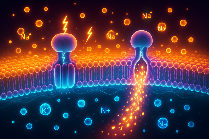
If depolarization matters, the ion channels that determine voltage should be relevant to cancer. They are. Potassium channels. K+ channels hyperpolarize cells (make Vmem more negative). Decreased K+ channel activity = depolarization = more cancerous phenotype. Many tumors show reduced expression of hyperpolarizing K+ channels. Sodium channels. Na+ channels depolarize cells. Certain voltage-gated sodium channels (particularly Nav1.5) are upregulated in metastatic cancers—breast, prostate, colon. Their activity correlates with invasiveness. Chloride channels. Cl- channels affect both voltage and cell volume. Altered Cl- channel expression is common in cancer and affects both proliferation and migration. Calcium channels. Ca2+ is a crucial second messenger. Ca2+ channel alterations in cancer affect signaling cascades that regulate proliferation, apoptosis, and invasion. Ion channels aren't just correlated with cancer. They're being explored as therapeutic targets. Several existing drugs that coincidentally block ion channels have anti-cancer effects. Drug companies are developing specific ion channel modulators for oncology.
Gap Junctions: The Tumor Suppressor Connection
Gap junctions connect adjacent cells, allowing small molecules and ions to flow between them. This creates electrical coupling—connected cells share voltage information. Gap junctions are tumor suppressors. The evidence is overwhelming. Loss of gap junction proteins (connexins) is one of the most common features of cancer. Tumor cells have fewer and less functional gap junctions than normal tissue. Restoration suppresses malignancy. When gap junction function is restored in cancer cells (by expressing connexins), the cells become less malignant—they grow slower, form fewer tumors, look more normal. Mechanism: Gap junctions allow a cell to "feel" its neighbors electrically. A normal cell that starts to depolarize experiences pull from its neighbors, whose negative potential helps restore normal voltage. A cancer cell that's lost gap junctions is electrically isolated—it can drift toward depolarization without correction. Gap junctions also allow the flow of tumor suppressors, growth inhibitors, and cell death signals between cells. An isolated cell doesn't receive the "stop growing" or "die" signals that coordinate normal tissue. Cancer isn't just cells growing wrong. It's cells that have stopped listening to their neighbors.
Tumor Microenvironment as Bioelectric Chaos
A tumor isn't just cancer cells. It includes surrounding stromal cells, immune cells, blood vessels, and extracellular matrix—the tumor microenvironment. The bioelectric environment of a tumor is disrupted at multiple levels: The cancer cells themselves are depolarized and electrically isolated from each other and from surrounding normal tissue. The surrounding tissue is remodeled. Altered pH (tumors are more acidic), changed ion concentrations, modified extracellular matrix—all affect bioelectric properties. Tissue architecture is disrupted. Gap junction networks that coordinate large tissue regions are fragmented. The tumor is a region of bioelectric incoherence in otherwise coherent tissue. This matters because the tumor microenvironment strongly influences cancer progression. Treatments that only target cancer cell genetics, while ignoring the corrupted environment, may miss critical drivers of malignancy.
The Normalization Hypothesis
Here's a therapeutic hypothesis: what if you could restore normal bioelectric patterns to cancerous tissue? This has been tested, with intriguing results: In vitro studies. Artificially hyperpolarizing cancer cells (making their Vmem more negative, more like normal cells) reduces proliferation, increases differentiation markers, and decreases invasive behavior. The cells become more "normal" when their voltage becomes more normal. In vivo studies. Modulating ion channels in tumor-bearing animals can affect tumor growth and metastasis. The effects aren't miraculous cures, but they're measurable. Tumor reversion. In some model systems, cancer cells placed in a normal tissue environment—surrounded by electrically normal cells—can revert toward normal phenotype. The tissue "talks sense" into them. This is the normalization hypothesis: cancer isn't just uncontrolled growth that must be killed. It's a loss of normal tissue coordination that might, in some cases, be restored. The bioelectric framework offers specific targets for this restoration.
Clinical Implications
Ion channel drugs. Several ion channel modulators are in cancer clinical trials. Some are repurposed drugs originally developed for other conditions (cardiac, neurological). The idea is to shift the bioelectric environment toward normal. Gap junction modulators. Drugs that enhance gap junction function could help restore tissue-level coordination. This approach is earlier-stage than ion channel targeting. Combination with conventional therapy. Bioelectric modulation might sensitize tumors to chemotherapy or radiation. A more "normal" bioelectric state might restore cell death pathways that malignancy suppresses. Early detection. Bioelectric abnormalities may precede visible tumors. Techniques that detect voltage changes could enable earlier cancer detection—before masses form. Prevention. If sustained depolarization promotes malignancy, maintaining normal bioelectric state might be protective. This is speculative but consistent with the framework. None of this replaces the genetic model. Mutations matter; oncogenes matter; tumor suppressors matter. The bioelectric framework adds a layer—an additional axis of intervention that operates through voltage and tissue coordination rather than cell-autonomous genetic targeting.
The Coherence Frame
Throughout this series, we've used "coherence" as a lens. Cancer fits this frame perfectly. Normal tissue is bioelectrically coherent. Cells maintain appropriate voltages. Gap junctions coordinate signals across tissue regions. Local voltage perturbations are corrected by coupling to neighbors. The system maintains itself. Cancer is bioelectric incoherence. Cells are depolarized and isolated. Gap junction networks are fragmented. Tissue-level coordination fails. The system no longer maintains its pattern. Coherence isn't metaphor here—it's measurable. You can map the voltage patterns, quantify the gap junction coupling, assess the tissue integration. And where that coherence breaks down, cancer emerges. This suggests that cancer prevention and treatment might benefit from a "coherence maintenance" perspective: not just killing aberrant cells, but maintaining the tissue-level patterns that keep cells behaving normally.
What We Don't Know
The bioelectric cancer framework is promising but not proven: Correlation vs. causation remains unclear for many observations. Depolarization accompanies cancer; whether it drives cancer in humans (beyond cell culture and animal models) is less certain. Specificity is a challenge. Ion channels exist throughout the body. Drugs that modulate voltage could have widespread effects. Finding cancer-specific interventions is non-trivial. Heterogeneity complicates everything. Different cancers, different patients, different bioelectric profiles. A one-size-fits-all bioelectric intervention seems unlikely. Integration with genetics is incomplete. We know oncogenes affect ion channels. The full map of how genetic alterations produce bioelectric changes—and how bioelectric changes influence genomic stability—is still being drawn. The framework is real. The therapeutic applications are still emerging.
Further Reading
- Levin, M. (2021). "Bioelectric signaling: Reprogrammable circuits underlying embryogenesis, regeneration, and cancer." Cell.
- Lobikin, M. et al. (2012). "Resting potential, oncogene-induced tumorigenesis, and metastasis: the bioelectric basis of cancer in vivo." Physical Biology.
- Mesnil, M. (2020). "Connexins and cancer: what can we learn from human disorders?" Cell Communication and Signaling.
- Prevarskaya, N., Skryma, R., & Shuba, Y. (2018). "Ion channels in cancer: are cancer hallmarks oncochannelopathies?" Physiological Reviews.
This is Part 6 of the Bioelectric Code series. Next: "The Bioelectric Future: A Synthesis."


Comments ()