Ion Channels and Gap Junctions: The Bioelectric Hardware
Before brains existed, cells were already talking electrically. Ion channels gate charged ions across membranes; gap junctions let cells share electrical state directly — the hardware layer of bioelectric intelligence.
Ion Channels and Gap Junctions: The Bioelectric Hardware
Series: Bioelectric Code | Part: 3 of 7 Primary Tag: FRONTIER SCIENCE Keywords: ion channels, gap junctions, connexins, bioelectric signaling, cell communication
Ion Channels: The Gates
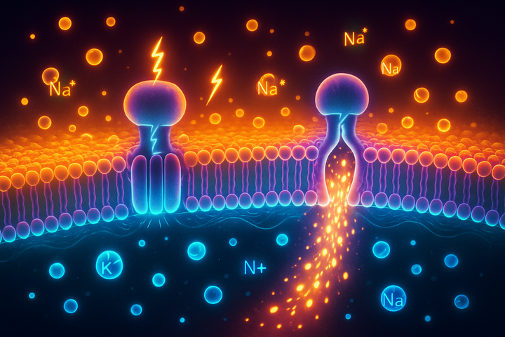
Ion channels are proteins that form pores in cell membranes, allowing specific ions to flow through. They're extraordinarily selective—a sodium channel lets sodium through but blocks potassium, despite the two ions differing by only 0.4 angstroms in radius. Types by gating mechanism: Voltage-gated channels: Open or close depending on membrane voltage. These create feedback loops—the voltage affects the channels which affect the ion flow which affects the voltage. Voltage-gated sodium, potassium, and calcium channels are the basis of action potentials. Ligand-gated channels: Open when a specific molecule binds. Neurotransmitter receptors (nicotinic acetylcholine receptors, GABA receptors) are examples. This links chemical signaling to electrical signaling. Mechanosensitive channels: Open in response to membrane stretch or pressure. Important in touch sensation and also in developmental signals from tissue mechanics. Leak channels: Constitutively open, providing baseline conductance that contributes to resting potential. Types by ion: Each major ion has dedicated channels:
- Sodium channels (Nav): Primarily known from action potentials but present in non-excitable cells too
- Potassium channels (Kv, Kir, etc.): The largest ion channel family; critical for setting resting potential
- Calcium channels (Cav): Calcium is a critical signaling ion; its entry triggers countless downstream events
- Chloride channels (ClC, CFTR): Affect cell volume, pH, and membrane potential
The diversity is staggering. The human genome encodes over 400 ion channel and transporter proteins. Each cell type expresses a specific repertoire that determines its electrical signature.
Gap Junctions: The Connections
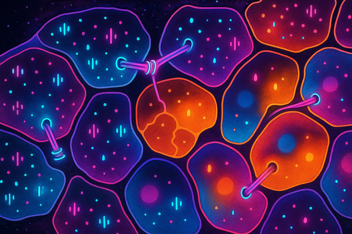
If ion channels control individual cells, gap junctions connect them. Gap junctions are protein complexes that create direct cytoplasmic channels between adjacent cells. Structure: A gap junction is made of two hemichannels (connexons), one contributed by each cell. Each hemichannel is made of six connexin proteins arranged in a ring. The joined hemichannels form a pore roughly 1.5 nm in diameter. What flows through: Small molecules up to ~1000 daltons can pass—ions, metabolites, signaling molecules like IP3 and cAMP. Not proteins or nucleic acids—they're too large. Electrical coupling: Because ions can flow through gap junctions, electrically coupled cells share membrane potential. If one cell depolarizes, current flows through gap junctions to neighbors, spreading the voltage change. This is how cardiac muscle synchronizes—gap junctions couple heart cells into a coordinated electrical syncytium. Selectivity: Different connexins form gap junctions with different properties—some more permeable to anions, others to cations. The specific connexins expressed determine which cells are coupled and what signals pass between them. Regulation: Gap junctions can be gated—closed by low pH, high calcium, phosphorylation, or voltage. This allows dynamic control of electrical coupling.
The Gap Junction Network
In tissues, gap junction connectivity forms a network topology. Not all cells are equally connected. Some are highly coupled; others are isolated. The topology matters: Compartmentalization: Groups of cells can be electrically isolated from each other by lack of gap junctions, allowing different Vmem patterns in different regions. Synchronization: Within a coupled region, gap junctions coordinate voltage. This is essential for heart rhythm, for synchronized calcium waves in development, for collective behavior. Long-range communication: Signals can propagate through gap junction networks across many cell diameters. A voltage change in one location can influence distant cells if they're gap-junctionally connected. The gap junction network is a communication infrastructure. Its topology shapes what information flows where.
Channelopathies: When Hardware Fails
Mutations in ion channels cause disease—channelopathies: Cardiac arrhythmias: Long QT syndrome, Brugada syndrome, and other conditions result from mutations in cardiac ion channels. The heart's electrical system misfires. Epilepsy: Some forms result from neuronal ion channel mutations affecting excitability. Myotonia: Skeletal muscle channel mutations causing muscle stiffness. Cystic fibrosis: A chloride channel (CFTR) mutation affecting epithelial function. These diseases demonstrate the importance of ion channels: disrupting them causes system-level dysfunction. Similarly, connexin mutations cause diseases: Deafness: Connexin 26 (GJB2) mutations are the most common cause of hereditary deafness. Gap junctions are essential for cochlear function. Skin diseases: Certain connexin mutations cause keratinization disorders. Cataracts: Lens connexins are critical for lens clarity. Cardiac defects: Connexin 43 mutations affect heart development and function. The hardware defects reveal the hardware's importance.
Developmental Patterning Through Channels
During development, specific ion channels and gap junctions are expressed in precise patterns, creating the bioelectric templates that guide morphogenesis. Left-right asymmetry: In early embryos, gap junction connections between cells on the left and right sides allow ion and signaling molecule flow that helps establish which side is which. Perturbing these connections disrupts organ asymmetry. Regeneration signaling: In planaria and other regenerators, specific ion channels are required for correct regeneration. Block them, and regeneration fails or produces abnormal outcomes. Organ boundaries: Gap junction expression often marks organ boundaries—cells within an organ are coupled; cells across boundaries are not. This electrical isolation may help maintain distinct organ identities. The molecular players (which channels, which connexins) vary by organism and tissue. But the principle is general: bioelectric hardware creates the patterns that guide development.
Tools for Manipulating the Hardware
Researchers can now precisely control bioelectric states: Pharmacology: Drugs targeting specific channels can hyperpolarize or depolarize cells. The antidiabetic drug metformin, for instance, affects AMPK and can influence membrane potential. Ivermectin affects chloride channels. Optogenetics: Light-sensitive channels (channelrhodopsins, halorhodopsins) can be expressed in specific cells, allowing light to control their voltage with millisecond precision. Chemogenetics: Designer receptors activated by designer drugs (DREADDs) allow chemical control of specific cell populations. Genetic manipulation: Overexpressing or knocking out specific channels in model organisms reveals their roles. CRISPR makes this increasingly precise. These tools allow causal experiments: change the channel expression, measure the bioelectric change, observe the biological consequence.
The Computational View
Ion channels and gap junctions form a bioelectric circuit. Like electronic circuits, bioelectric circuits can compute: Memory: Voltage-gated channels create positive feedback loops that can hold states. A depolarization can be self-sustaining if it opens channels that maintain the depolarization. Oscillation: Channel kinetics can create oscillators—cells that rhythmically depolarize and repolarize. The heart's pacemaker cells are an example. Pattern formation: Reaction-diffusion dynamics, where voltage affects channel activity affects ion flow affects voltage, can create spatial patterns—stripes, spots, gradients. Decision-making: Threshold effects in voltage-gated channels create binary decisions. Above threshold, the channel opens; below, it stays closed. Tissues can make "decisions" based on reaching voltage thresholds. This computational capacity is why bioelectric patterns can encode complex anatomical information. The hardware is simple (channels and junctions), but the network dynamics are rich enough to support sophisticated pattern formation and memory.
Further Reading
- Hille, B. (2001). Ion Channels of Excitable Membranes. 3rd ed. Sinauer Associates.
- Goodenough, D.A. & Paul, D.L. (2009). "Gap junctions." Cold Spring Harbor Perspectives in Biology.
- Levin, M. & Martyniuk, C.J. (2018). "The bioelectric code: An ancient computational medium for dynamic control of growth and form." BioSystems.
This is Part 3 of the Bioelectric Code series. Next: "Morphogenetic Fields: How Bodies Know Their Shape."


Comments ()