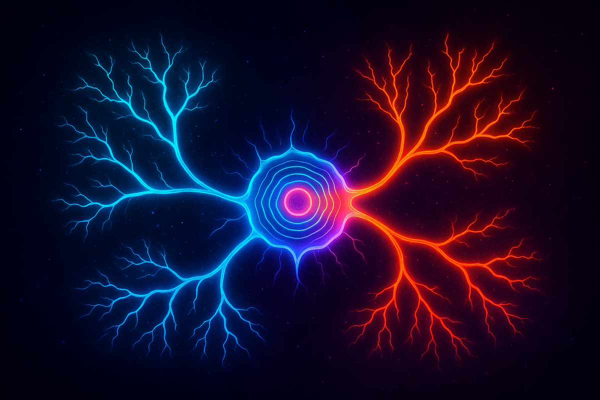
The Voltage of Life: Membrane Potential and Cell Fate
Before a cell divides or differentiates, it checks its voltage. Membrane potential (Vmem) is a signaling layer that sits above the genome and shapes what a cell becomes.
The Voltage of Life: Membrane Potential and Cell Fate
Series: Bioelectric Code | Part: 2 of 7 Primary Tag: FRONTIER SCIENCE Keywords: membrane potential, Vmem, depolarization, hyperpolarization, cell fate, differentiation
What Vmem Tells a Cell
The membrane potential isn't just a passive consequence of ion gradients—it's a signal. Cells read their own voltage and respond accordingly. Depolarized cells (less negative, e.g., -20 mV):
- Tend toward proliferation
- Resist differentiation
- Characteristic of stem cells, cancer cells, embryonic cells
Hyperpolarized cells (more negative, e.g., -70 mV):
- Tend toward quiescence
- Favor differentiation
- Characteristic of mature, differentiated tissue
This isn't just correlation. Experimentally manipulating Vmem changes cell behavior:
- Force depolarization in differentiated cells → they can re-enter the cell cycle
- Force hyperpolarization in proliferating cells → promotes differentiation, slows division
- Block ion channels that maintain specific voltage states → disrupt developmental patterns
The voltage is causally involved in cell fate decisions.
How Voltage Affects Molecular Pathways
How does a 50-millivolt change translate into molecular consequences? Voltage-sensitive signaling:
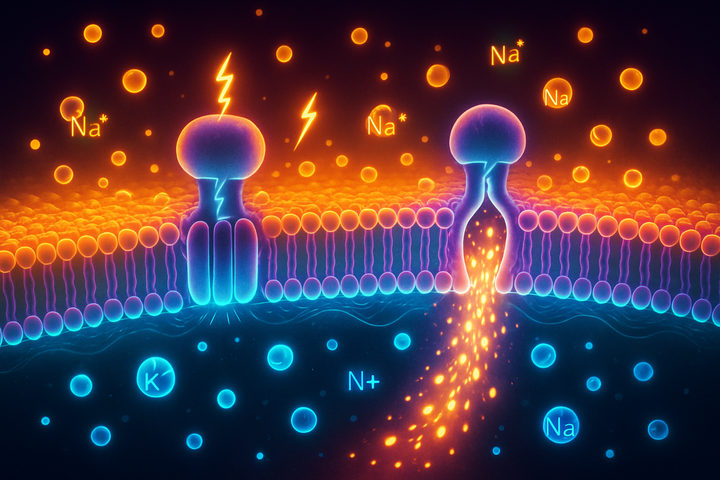
- Voltage-gated calcium channels open at specific voltages, allowing calcium influx. Calcium is a master second messenger affecting hundreds of downstream pathways.
- Voltage-sensitive phosphatases (VSPs) change their activity with voltage, affecting signaling cascades.
- The electrochemical gradient affects transport of signaling molecules.
Gene expression:
- Ion channel activity can ultimately affect transcription factors. NFAT, CREB, and others are regulated by calcium or other voltage-influenced pathways.
- Chromatin state can be affected by nuclear calcium or by metabolic changes driven by electrical activity.
Metabolic changes:
- Mitochondrial function is voltage-sensitive. The mitochondrial membrane potential affects ATP production.
- Metabolic intermediates feed into epigenetic modifications.
The bioelectric signal is transduced into biochemical signals, which affect gene expression, which affects cell behavior. Vmem is an input variable in a complex computation.
The Vmem Map of Development
During development, tissues show characteristic patterns of Vmem: Eye development in Xenopus (frog): The region that will become the eye shows a specific voltage signature. Disrupt it, and eye development fails. Reproduce that signature elsewhere, and you can induce eyes in abnormal locations. Limb regeneration in salamanders: The regeneration blastema (the mass of cells that will regrow the limb) has a distinct bioelectric state. Blocking certain ion channels prevents regeneration. Activating the right channels can enhance it. Left-right asymmetry: Before anatomical asymmetry is visible, there are voltage differences between left and right sides of the embryo. Ion channel mutations that disrupt this pattern cause situs inversus (reversed organ positions) or heterotaxy (mixed-up organs). The Vmem pattern is a pre-pattern—information about tissue identity encoded before morphology is visible.
Cancer: The Voltage Connection
Multiple lines of evidence connect Vmem to cancer: Tumor cells are depolarized: Across cancer types, malignant cells tend to have less negative resting potentials than their normal counterparts. This was observed decades ago but is now being systematically characterized. Depolarization promotes malignant behaviors: Experimentally depolarizing normal cells can induce some cancer-like behaviors (increased proliferation, resistance to apoptosis). Hyperpolarizing cancer cells can suppress malignancy in some contexts. Ion channel expression changes in cancer: Many cancers show altered expression of specific ion channels. Some of these channels are being explored as drug targets. Bioelectric normalization: In some experiments, restoring normal voltage patterns to cancerous tissue has reduced tumor growth or caused tumor cells to behave more normally. The strong version of the hypothesis: cancer is partly a bioelectric disease—a failure to maintain the voltage state that normally keeps cells quiescent and differentiated. This doesn't mean cancer isn't genetic. Genetic mutations in oncogenes and tumor suppressors are real and important. But bioelectric signals may be a layer that interprets and modifies how genetic changes manifest. It's another control system, disrupted in malignancy.
Measuring and Manipulating Vmem
Measurement tools:
- Patch-clamp electrophysiology: Gold standard, but invasive and low throughput
- Voltage-sensitive dyes: Fluorescent molecules that change brightness with voltage. Allows imaging of voltage patterns across tissues.
- Genetically encoded voltage indicators (GEVIs): Protein sensors that can be expressed in specific cell types, allowing voltage imaging in living animals.
Manipulation tools:
- Channel blockers/openers: Drugs that affect specific ion channels, changing the conductances that determine Vmem
- Optogenetics: Light-sensitive ion channels that allow precise, rapid control of voltage with light
- Ion channel misexpression: Genetically introducing channels from other species (e.g., expressing a hyperpolarizing channel in normally depolarized tissue)
These tools are enabling the systematic study of bioelectric control. We can now read and write the bioelectric code in experimental settings.
Why Vmem Is Information
Here's the conceptual shift: membrane potential isn't just a cell's electrical state. It's information that cells use to make decisions. Positional information: Vmem patterns tell cells where they are in a tissue. High-Vmem region? You're in the future eye field. Low-Vmem region? Different fate. State information: Vmem tells a cell about its own status. Differentiated and quiescent? You should be hyperpolarized. Proliferating? Depolarized is expected. Communication: Voltage gradients allow cells to sense and respond to their neighbors. A cell surrounded by depolarized neighbors receives different signals than one in hyperpolarized tissue. This information view is crucial. Vmem isn't just physics happening to cells—it's a signal that cells are evolved to interpret.
The Evolutionary Perspective
Bioelectric signaling is ancient. Even bacteria have ion channels and membrane potentials. The basic machinery predates neurons by billions of years. Evolution co-opted this pre-existing electrical system for multiple purposes: 1. Basic homeostasis: Regulating cell volume, pH, nutrient transport 2. Developmental signaling: Coordinating cell behavior across tissues 3. Regeneration: Encoding pattern information for repair 4. Nervous system: Elaborating the basic system into rapid, long-distance communication Neurons didn't invent bioelectricity; they specialized it. The widespread bioelectric properties of non-neural cells are the ancestral condition.
The Coherence View
Vmem is a coherence variable. When tissue Vmem patterns are organized—appropriate gradients, appropriate values for each cell type—the tissue develops and functions normally. The pattern constitutes a kind of distributed memory about what the tissue should be. Cancer is incoherence: cells with aberrant voltage, failing to read and respond to normal bioelectric signals. Regeneration is coherence restoration: re-establishing the bioelectric pattern that encodes the correct anatomical structure. Understanding Vmem as a coherence variable suggests therapeutic strategies: don't just target genes or proteins—target the bioelectric state that coordinates cell behavior.
Further Reading
- Blackiston, D.J. et al. (2009). "Bioelectric controls of cell proliferation." Cell Cycle.
- Yang, M. & Bhavsar, M. (2016). "Voltage-dependent processes in cells and the role of ion channels." Physiological Reviews.
- Levin, M. (2014). "Endogenous bioelectrical networks store non-genetic patterning information during development and regeneration." Journal of Physiology.
This is Part 2 of the Bioelectric Code series. Next: "Ion Channels and Gap Junctions: The Hardware."


Comments ()