Morphogenetic Fields: How Bodies Know Their Shape
Genes are the recipe, but something else sets the table. Morphogenetic fields — gradients of chemical signals and bioelectric patterns — tell each cell where it is and what it should become. The body's shape is information, stored outside the genome.
Morphogenetic Fields: How Bodies Know Their Shape
Series: Bioelectric Code | Part: 4 of 7 Primary Tag: FRONTIER SCIENCE Keywords: morphogenetic fields, pattern formation, bioelectric gradients, positional information, developmental biology
The Positional Information Problem
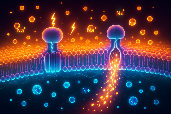
Each cell in your body contains the same genome. Yet cells in your liver do liver things, cells in your brain do brain things, cells in your fingers form fingers. How do cells know where they are? Classical answer: chemical gradients. Morphogens—signaling molecules like Sonic Hedgehog, BMPs, Wnts—form concentration gradients across developing tissues. Cells read the local morphogen concentration and respond accordingly. High Shh concentration means "become ventral"; low means "become dorsal." This is real and important. But it's not the whole story. The bioelectric answer: voltage patterns. Before morphogen gradients are established, bioelectric gradients exist. Voltage patterns can provide positional information earlier and at larger scales. They complement and interact with chemical gradients.
Evidence for Bioelectric Morphogenetic Fields
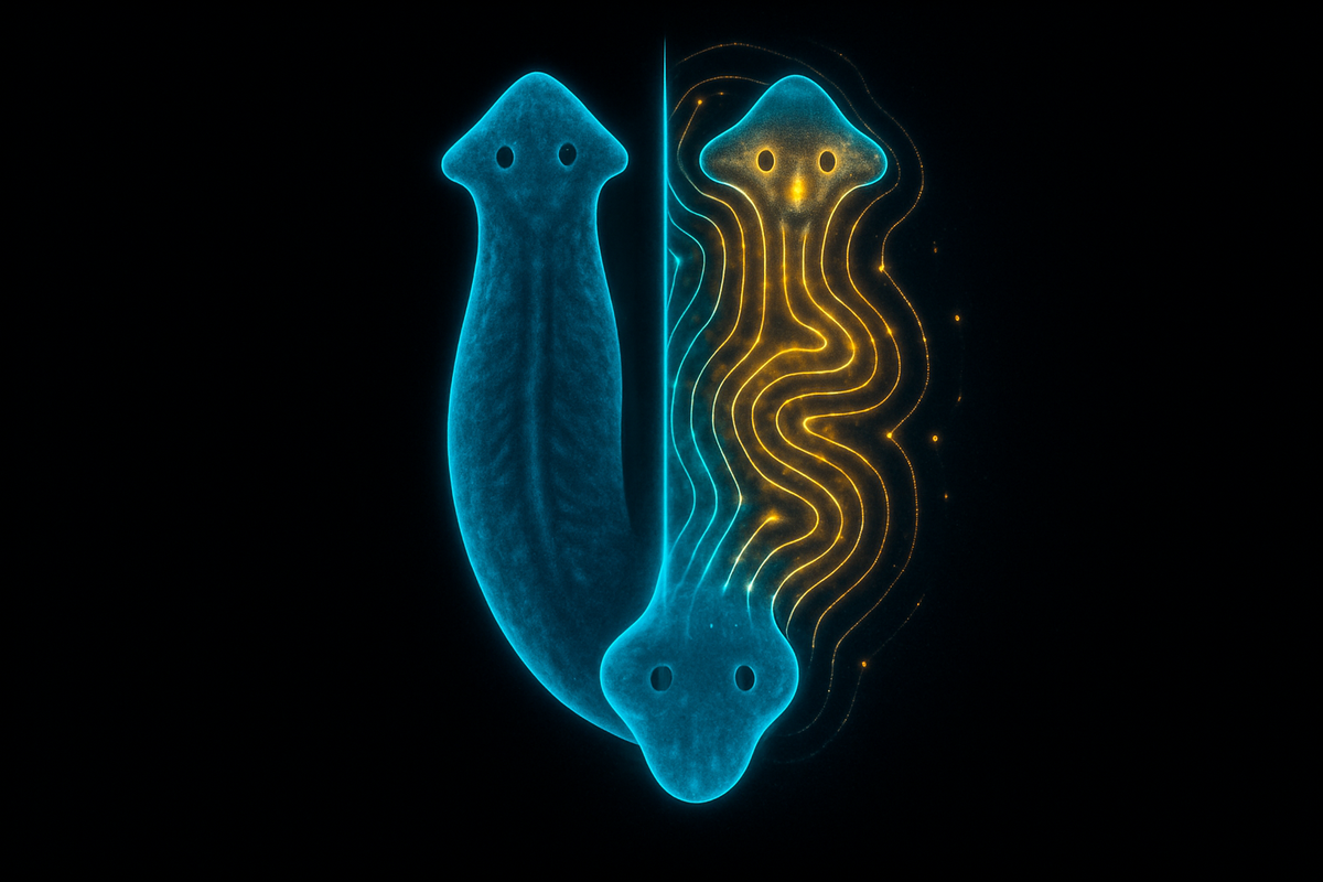
Regeneration in planaria: Planarian flatworms can regenerate from tiny fragments. Cut a worm in half; each half regenerates the missing parts. How does a fragment "know" which end should grow a head and which a tail? Michael Levin's lab showed that bioelectric gradients encode this polarity. The head region has a different voltage than the tail. Block specific ion channels, and you can create two-headed worms—the voltage cue that says "grow tail here" is disrupted. Crucially, once you've changed the bioelectric pattern, the effect persists through subsequent rounds of regeneration. The worm "remembers" its new polarity in its bioelectric state. Eye induction in Xenopus: Normally, eyes develop in specific head locations. Levin's lab showed that expressing certain ion channels in other locations—the gut, the tail—could induce functional eyes to form there. The ion channel manipulation creates a bioelectric signature that cells interpret as "eye field." The cells respond by making eyes, even in aberrant locations. Limb regeneration signaling: In amphibians that regenerate limbs, the regeneration blastema has a characteristic bioelectric state. Manipulating this state affects regeneration. Adding depolarizing factors can trigger regeneration-like responses in tissues that don't normally regenerate.
How Bioelectric Fields Encode Pattern
The bioelectric field is a spatial pattern of voltage across tissue. How does this encode anatomical information? Gradients provide positional information. If voltage smoothly varies from head to tail, cells can read their position in the gradient. "My voltage is X, which means I'm Y% of the way along the axis." Boundaries define regions. Sharp transitions in voltage can mark organ boundaries. Cells on either side of a voltage boundary know they're in different regions. Dynamic patterns coordinate timing. Bioelectric patterns can change over developmental time, providing information not just about position but about developmental stage. Feedback creates stability. Voltage affects gene expression, which affects ion channel expression, which affects voltage. These feedback loops can stabilize patterns, creating robust developmental trajectories. The field is continuously computed by the cells themselves—it's a distributed, self-organizing pattern that emerges from cellular interactions.
The Bioelectric-Genetic Interface
Bioelectric fields don't replace genetic programs; they interact with them. Gene expression responds to voltage: Many developmental genes are influenced by voltage-sensitive pathways. Changing Vmem can change what genes cells express. Genes encode bioelectric machinery: The ion channels and gap junctions that create bioelectric patterns are genetically encoded. Development sets up the bioelectric pattern by controlling which channels are expressed where. Epigenetic integration: Bioelectric signals can affect chromatin state, potentially creating lasting changes in gene expression. The bioelectric pattern becomes part of the epigenetic landscape. The relationship is bidirectional: genes shape bioelectric patterns; bioelectric patterns shape gene expression. They're not separate systems but interacting layers of developmental control.
Morphogenetic Field Memory
One of the most striking findings: bioelectric patterns can store information that persists. In planaria, if you manipulate the bioelectric pattern to create a two-headed worm, that worm will regenerate as two-headed even after the original manipulation is long gone. The abnormal pattern is remembered. This suggests the bioelectric field acts as a kind of pattern memory—storing information about what the target morphology should be. The pattern isn't just an instruction; it's a stable state that gets re-established. This is reminiscent of computer memory. Bits persist until they're actively changed. Bioelectric patterns persist until something resets them.
Clinical Implications
If bioelectric fields guide development and regeneration: Birth defects might be addressable by correcting bioelectric patterns. Instead of (or in addition to) gene therapy, voltage manipulation might restore normal development. Regenerative medicine might trigger regeneration by establishing the right bioelectric cues. If we knew the voltage pattern for "grow a finger here," we might be able to induce it. Cancer treatment might target bioelectric abnormalities. If tumors have disrupted voltage patterns, restoring normal patterns might suppress malignancy. These are hypotheses being tested. The clinical applications are not yet realized, but the scientific basis is being established.
The Coherence Interpretation
Morphogenetic fields are coherence patterns. They represent the organized state a tissue "should" be in. When the bioelectric field is coherent—the right gradients, the right boundaries, the right dynamic coordination—development proceeds normally. When the field is disrupted—wrong voltage values, broken boundaries, loss of coordination—development goes awry: birth defects, failed regeneration, possibly cancer. Coherence is maintained by the same mechanisms that create it: ion channels, gap junctions, and their feedback interactions. The system self-organizes toward coherent states. The morphogenetic field concept, once dismissed as mystical, now has a concrete interpretation: it's the spatial pattern of Vmem and its associated molecular state, computed continuously by cellular interactions, encoding positional information that guides cell behavior.
Further Reading
- Levin, M. (2012). "Morphogenetic fields in embryogenesis, regeneration, and cancer: Non-local control of complex patterning." BioSystems.
- Adams, D.S. & Levin, M. (2013). "Endogenous voltage gradients as mediators of cell-cell communication." Developmental Biology.
- Mathews, J. & Bhavsar, M. (2021). "The bioelectric code: A unified framework for understanding morphogenesis and regeneration." Current Opinion in Cell Biology.
This is Part 4 of the Bioelectric Code series. Next: "Planarian Regeneration: The Champion Regenerators."


Comments ()