Optogenetics: Controlling Neurons with Light
Before optogenetics, neuroscientists could record neurons or stimulate brain regions bluntly with electricity. Deisseroth's method uses channelrhodopsins to turn specific cell types on or off with a flash of light — giving researchers causal control over circuits for the first time.
Optogenetics: Controlling Neurons with Light
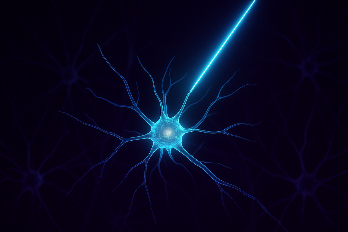
In 2005, Karl Deisseroth's lab at Stanford did something that sounded impossible. They took a gene from algae—a gene that codes for a light-sensitive protein—and inserted it into mammalian neurons. When they shone blue light on those neurons, they fired. When they turned off the light, they stopped. On. Off. Like a switch. This was not how neuroscience had worked before. Before optogenetics, we could observe the brain. We could measure activity, record from neurons, watch patterns unfold. But intervening was crude—electrodes that stimulated entire regions, drugs that affected the whole brain, lesions that destroyed tissue. Correlation was easy. Causation was hard. Optogenetics changed the rules. Now you could activate one specific cell type, in one specific region, at one specific moment, with millisecond precision. You could ask: what happens if I turn on exactly these neurons? And you could get an answer. Optogenetics didn't just give neuroscience a new tool. It shifted the field from watching brains to controlling them.
The Technical Setup (How It Actually Works)
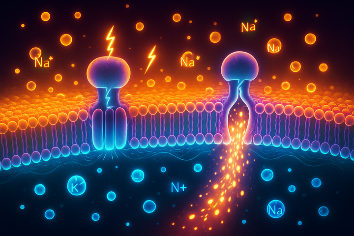
Getting light-sensitive proteins into the right neurons requires genetic manipulation. Here's the typical workflow: Step 1: Choose your target cell type. You don't want to activate all neurons—you want to activate specific types. Maybe you're interested in dopamine neurons in the reward circuit. Maybe inhibitory interneurons in the cortex. Each cell type expresses characteristic genes. Step 2: Design a viral vector. You take a virus—usually an adeno-associated virus (AAV), which is safe and well-studied—and engineer it to carry the channelrhodopsin gene along with a promoter that's only active in your target cell type. When the virus infects neurons, only the target cells will express the light-sensitive protein. Step 3: Inject the virus. You use stereotactic surgery to inject the viral vector into a precise brain region. Over a few weeks, the target neurons start producing channelrhodopsin and inserting it into their membranes. Step 4: Implant a fiber optic. To deliver light deep in the brain, you surgically implant a thin fiber optic cable. The tip sits right next to the neurons you want to control. Step 5: Turn on the light. Blue light travels through the fiber optic, hits the channelrhodopsin-expressing neurons, and they fire. You control the timing, the duration, the intensity. Millisecond precision. It sounds invasive, and it is—optogenetics is mostly used in animal research, not humans. But the precision is extraordinary. You can stimulate one cell type while leaving neighboring cells untouched. You can turn cells on during one phase of behavior and off during another. You can ask causal questions that were previously unanswerable.
The Discoveries That Followed
Within a decade, optogenetics had rewritten textbooks on multiple brain systems.
Memory: Making and Erasing
Susumu Tonegawa's lab at MIT used optogenetics to identify "memory engram cells"—the specific neurons that encode a particular memory. They genetically labeled neurons that were active while a mouse learned to fear a specific environment. Later, when those same neurons were optogenetically reactivated—even in a completely different environment—the mouse froze in fear. The memory was stored in those specific cells, and activating them was sufficient to recall it. More strikingly, they could create false memories. They activated fear-encoding neurons while the mouse was in a safe environment, and the mouse later feared that safe environment—even though nothing bad had ever happened there. Optogenetics proved that memories live in specific neurons and that activating those neurons is sufficient to trigger recall.
Reward: The Dopamine Circuit
Dopamine neurons in the midbrain have long been associated with reward and motivation. But correlation isn't causation. Just because dopamine neurons fire when an animal gets a reward doesn't prove they cause the experience of reward. With optogenetics, researchers could directly activate dopamine neurons. Animals would work obsessively for stimulation—pressing a lever over and over to turn on the light that activated their dopamine cells. This confirmed that dopamine neuron activity is sufficient to produce reward-like motivation. But the story is more nuanced than "dopamine = pleasure." Optogenetic experiments helped reveal that dopamine signals prediction error—the difference between expected and actual reward. Dopamine neurons fire not for reward itself, but for unexpected reward. This precision was only possible because optogenetics could deliver stimulation at exact moments, testing specific hypotheses about timing.
Sleep: Switching Brain States
Sleep researchers used optogenetics to identify neurons that can switch the brain between states. Activating certain neurons in the hypothalamus could instantly put an animal to sleep. Activating others could wake it up. This wasn't just observation—it was control. The existence of these "switch neurons" had been hypothesized, but optogenetics proved they existed and characterized exactly which cell types were responsible.
Parkinson's: Circuit Dysfunction
Parkinson's disease involves the death of dopamine neurons and dysfunction of motor circuits. Deep brain stimulation (DBS)—electrodes that deliver current to specific brain regions—can alleviate symptoms, but we didn't fully understand why it worked. Optogenetic experiments in mice revealed that the therapeutic effect likely comes from stimulating specific axons passing through the target region, not the region's neurons directly. This changed understanding of how DBS works and suggested ways to improve it.
The Precision Revolution
Before optogenetics, neuroscience was stuck in a dilemma. You could observe with high precision—recording from individual neurons, imaging activity patterns, measuring timing down to milliseconds. But you could only intervene crudely—lesioning whole brain areas, injecting drugs that affected everything, using electrodes that stimulated all neurons in range. The mismatch was devastating for causal inference. If you lesion a region and behavior changes, you know the region was involved—but you don't know which cell types, which projections, which temporal patterns mattered. Optogenetics matched the precision of intervention to the precision of observation.
- Want to know if a specific cell type is necessary for a behavior? Silence just those cells during the behavior.
- Want to know if a specific timing pattern matters? Stimulate with that exact pattern and see if the behavior follows.
- Want to know if a projection from region A to region B is causal? Stimulate axon terminals of A neurons in region B specifically.
This isn't just a better screwdriver. It's a fundamentally different kind of science—circuit-level causality, where you can test exactly which components of a system are doing what.
Limitations and Challenges
Optogenetics is powerful but not magic. Invasive surgery. You need to inject viruses and implant fiber optics. This limits use in humans to clinical situations where the brain is already being accessed surgically (like epilepsy treatment). Not all neurons are accessible. Some brain regions are hard to target. Some cell types don't have clean genetic markers. The technique works best for well-defined populations with known gene expression patterns. It's not natural activity. Optogenetic stimulation produces synchronized activation of many neurons simultaneously—different from natural activity patterns where neurons fire more heterogeneously. This can produce unnaturally strong effects or miss the nuances of real neural coding. Expression variability. Not all target neurons express the opsin at the same level. Some may express a lot, some a little, some none. This creates variability in the strength and distribution of stimulation. Light scatter. Brain tissue scatters light, so stimulation isn't perfectly localized. You can hit neurons near the fiber tip, but light spreads, meaning some non-target neurons may be affected. Despite these limitations, optogenetics remains the gold standard for causal intervention in neural circuits. When you read a paper saying "X neurons cause Y behavior," chances are optogenetics was involved.
The Human Frontier
Can optogenetics work in humans? The technical barriers are significant. You'd need to deliver viral vectors to the human brain (doable but risky), implant fiber optics (invasive), and ensure long-term safety of foreign proteins in human neurons (unknown). But the potential is tantalizing. Retinal diseases. The most advanced human optogenetics work targets the eye. In retinitis pigmentosa, photoreceptors die but other retinal cells remain. Researchers have given those surviving cells channelrhodopsin, effectively making them light-sensitive—bypassing the dead photoreceptors. Clinical trials are ongoing, and some patients have recovered functional vision. Brain diseases. Optogenetics could, in principle, offer more precise intervention than deep brain stimulation—targeting specific cell types rather than stimulating everything in range. For Parkinson's, epilepsy, or depression, this precision could mean better outcomes with fewer side effects. Human trials are years away, but the rationale is clear. Understanding psychiatric disorders. Even if optogenetics never becomes a human therapy, the animal research informs human medicine. By identifying exactly which circuits cause symptoms in animal models, we can develop better drugs, better stimulation protocols, and better diagnostic tools for humans.
The Legacy of a Light-Sensitive Algae Gene
Karl Deisseroth was trained as a psychiatrist. He started optogenetics because he was frustrated—frustrated that he could see patients suffering from depression, anxiety, schizophrenia, and he couldn't tell them what was actually going wrong in their brains. Psychiatric drugs work on the whole brain. Talk therapy works on the whole mind. Neither gives you circuit-level understanding. Neither tells you: this neuron type, in this region, firing at this time, is causing this symptom. Optogenetics was an attempt to build tools precise enough to ask those questions. Twenty years later, we still don't have complete answers. Psychiatric disorders are complex, and animal models have limitations. But we've learned more about reward circuits, fear circuits, social circuits, and sleep circuits in two decades of optogenetics than in the previous century. The gene from algae became the flashlight that lets us see inside the brain—and more importantly, the switch that lets us control what we find. Neuroscience went from watching to intervening. That's the optogenetics revolution.
Further Reading
- Deisseroth, K. (2011). "Optogenetics." Nature Methods. (The original review by the field's founder)
- Boyden, E.S. et al. (2005). "Millisecond-timescale, genetically targeted optical control of neural activity." Nature Neuroscience.
- Liu, X. et al. (2012). "Optogenetic stimulation of a hippocampal engram activates fear memory recall." Nature. (The false memory study)
- Deisseroth, K. (2021). Projections: A Story of Human Emotions. Random House. (Memoir connecting optogenetics to psychiatry)
This is Part 3 of the New Neuroscience series. Previous: "Connectomics: Mapping the Brain's Wiring." Next: "Glial Cells: The Other Half of Your Brain"—the support cells that might be more important than neurons.


Comments ()