Clinical TDA: Topological Biomarkers for Brain Disorders
Depression looks different on a topological map of brain activity. So does trauma. TDA — topological data analysis — extracts geometric features from neural data that standard connectivity measures miss, pointing toward objective biomarkers for conditions that currently lack them.
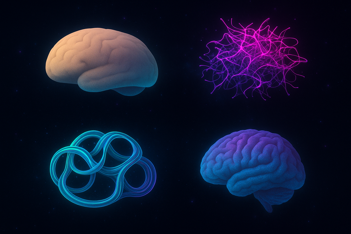
Clinical TDA: Topological Biomarkers for Brain Disorders
Series: Topological Data Analysis in Neuroscience | Part: 8 of 9
Psychiatry has a measurement problem.
We diagnose based on symptoms—clusters of behaviors, subjective reports, clinical observation. But symptoms don't respect clean categories. Depression bleeds into anxiety. Bipolar looks like ADHD during certain phases. Trauma produces symptoms that overlap half the diagnostic manual.
And critically: we have no objective markers. No blood test for depression. No brain scan that definitively diagnoses schizophrenia. No biomarker that tells you if treatment is working before waiting months to see if symptoms improve.
Until now, maybe.
Because topological data analysis is revealing geometric signatures of psychiatric and neurological disorders—measurable features in brain structure and function that correlate with diagnosis, predict treatment response, and track recovery. Objective, quantitative biomarkers based on the actual shape of neural dynamics.
Mental illness has a topology. And TDA can see it.
The Biomarker Promise
What makes a good biomarker?
1. Objective: Measurable without depending on subjective report.
2. Sensitive: Changes reliably with disease state.
3. Specific: Distinguishes between similar conditions.
4. Predictive: Forecasts outcomes, treatment response, progression.
5. Accessible: Obtainable with reasonable cost/effort in clinical settings.
Topological features from TDA can meet all five criteria:
They're objective—Betti numbers, persistence values, barcode diagrams are mathematical quantities extracted from brain imaging data. No interviewer bias, no self-report variance.
They're sensitive—as we've seen, topology changes dramatically with brain state. Disease alters geometry, and TDA detects those alterations.
They're specific—different disorders produce different topological disruptions. The geometric signature of depression differs from schizophrenia, from autism, from Alzheimer's.
They're predictive—baseline topology predicts who responds to treatment, who progresses to more severe stages, who recovers fully versus partially.
And increasingly, they're accessible—standard MRI protocols can provide the data. Computational pipelines extract the topology. No exotic equipment, no invasive procedures.
The question isn't whether topological biomarkers exist. It's how fast we can validate and deploy them.
Depression: Flattened Topology
Major depressive disorder shows one of the clearest topological signatures: dimensional collapse.
Healthy resting-state networks have rich topology—high Betti numbers in multiple dimensions, persistent features spanning many scales, complex geometric structure across default mode, salience, and central executive networks.
Depression flattens this.
Reduced β₁: Fewer loops in functional connectivity. The circuits that normally allow activity to circulate, that support self-referential thought and memory integration, are diminished.
Reduced β₂: Fewer voids. The high-dimensional cavities that characterize complex integration are collapsed. The manifold becomes simpler, lower-dimensional.
Shorter persistence: What topological features remain don't last. They appear at one threshold, disappear at slightly different threshold. Nothing robust, nothing stable.
Network-specific effects: Default mode network shows the most severe topological reduction. This is the network disrupted in depression phenomenologically—rumination, negative self-focus, memory bias all involve DMN dysfunction. The topology collapse is localized to precisely the system producing symptoms.
And critically: severity correlates with degree of collapse. Mild depression shows moderate topological reduction. Severe depression shows near-total flattening. Treatment response shows topological restoration—as symptoms improve, Betti numbers increase, persistence lengthens.
This isn't just correlation. The topology appears to be mechanistically relevant. Dimensional collapse means fewer accessible brain states. Depression feels like narrowed possibility—and geometrically, that's precisely what it is. The state-space has contracted. Fewer futures seem reachable because the manifold supporting future-oriented cognition has simplified.
Treatment restores dimensionality. Antidepressants, therapy, TMS—whatever works appears to work partly by rebuilding topological complexity.
Schizophrenia: Incoherent Complexity
If depression is too simple, schizophrenia is complex in the wrong ways.
Topological analysis reveals: excess local features without global integration.
β₀ (connected components) is abnormally high—the brain fragments into more separate pieces than healthy controls. Networks that should integrate are functioning independently.
β₁ shows bizarre patterns—many short-lived loops that don't organize into stable structures. The geometry is busy but incoherent.
Higher-dimensional features appear in wrong places. Regions that shouldn't show complex topology (primary sensory areas) develop high-dimensional structures. Regions that should integrate globally (prefrontal cortex) show impoverished topology.
Persistence distributions are abnormal. Instead of a few long-lived robust features and many short-lived noise features, schizophrenia shows intermediate persistence everywhere—nothing clearly signal, nothing clearly noise.
This matches phenomenology eerily well. Schizophrenia often involves overwhelmed integration—too much salience, too many connections, inability to filter noise from signal. Hallucinations might reflect aberrant topological features that create false perceptual structure. Delusions might be attempts to make sense of incoherent geometric organization that doesn't support stable models of reality.
And treatment? Antipsychotics appear to reduce topological complexity. They don't just sedate—they specifically reduce the incoherent high-dimensional features while preserving (or even enhancing) the robust low-dimensional integration.
Clozapine, the most effective antipsychotic, produces the most dramatic topological normalization. Less effective drugs show less geometric correction. Maybe potency correlates with topology-restoring power.
Alzheimer's Disease: Progressive Topological Degradation
Alzheimer's is neurodegeneration—progressive loss of neurons and synapses. As connectivity degrades, function collapses. TDA can track this geometrically.
Early stages (mild cognitive impairment): Subtle topological changes detectable before clinical diagnosis.
Reduced persistence in hippocampal-cortical loops. The geometry supporting memory encoding and retrieval shows weakening. Betti numbers remain near-normal, but features don't last as long.
Middle stages (moderate dementia): Clear dimensional collapse.
β₁ drops significantly. Loops disappear. Default mode network topology degrades. Higher-order features (β₂, β₃) are lost. The manifold flattens progressively.
Late stages (severe dementia): Near-total topological simplification.
Almost all higher-dimensional structure gone. Only basic connectivity (β₀) remains, and even that fragments. The geometric organization supporting integrated cognition has dissolved.
Progression rate is predictable from topology. Patients with faster topological decline progress faster clinically. Those who maintain more topology longer maintain function longer.
This suggests early intervention targets: Can we slow topological degradation? Exercise, cognitive stimulation, social engagement—interventions that seem to help all show effects on brain topology. They don't prevent degeneration, but they slow geometric collapse.
And potential treatment monitoring: Track Betti numbers over time. If they stabilize, treatment is working. If they continue declining, try different approaches.
Autism: Different, Not Deficient
Autism is not simple deficit. It's different neurodevelopmental organization. And topology reveals this clearly.
Autistic brains don't have universally reduced topology. They have different topology:
Higher local connectivity. More small loops, more local integration. Short-range connections form richer geometric structure.
Different long-range topology. Not universally reduced, but reorganized. Some long-range connections that are weak in neurotypical brains are strong in autistic brains, and vice versa.
Network-specific patterns. Visual networks often show more topological complexity in autism. Motor networks show different patterns. Social brain networks show reorganization, not simply reduction.
Heterogeneity. Autism is called a spectrum for a reason. Topological analysis reveals just how diverse autistic brain geometry actually is. There are multiple geometric subtypes—different patterns of reorganization that produce superficially similar behavioral presentations.
This reframes autism appropriately: not broken topology, but different coherence architecture. Different ways of organizing neural state space that produce different cognitive strengths and challenges.
Autistic individuals often excel at pattern recognition, systemizing, detail-oriented processing. Topologically, this might reflect richer local geometric structure—more dimensions available for fine-grained discrimination.
Challenges with social cognition and flexibility might reflect different global integration patterns—not absent, but organized such that certain state transitions are harder, certain intuitions less automatically accessible.
Support strategies should target topology: Help build bridges between local geometric richness and global integration. Scaffold the transitions between states that anatomy makes difficult. Don't flatten the topology to match neurotypical patterns—that loses the strengths along with addressing the challenges.
PTSD and Trauma: Topological Scars
Post-traumatic stress disorder shows fragmented, unstable topology consistent with its phenomenology.
Hyperconnectivity in fear circuits. Amygdala-based networks show excessive topological features—too many loops, over-integrated threat processing. The geometry that should activate during actual danger stays activated chronically.
Disconnection from regulation networks. Prefrontal-limbic loops that normally modulate emotional responses show reduced topology. The circuits that could dampen overactive fear aren't geometrically integrated enough to work effectively.
Stuck attractors. Certain topological configurations become too stable. The brain falls into traumatic memory states and can't escape—the manifold has deep basins that trap neural dynamics.
State transition rigidity. Difficulty moving between calm and alert states. The topological paths between these configurations are disrupted. What should be smooth transitions become jarring jumps.
Successful trauma therapy—whether EMDR, prolonged exposure, cognitive processing, or somatic approaches—appears to reshape trauma topology:
Reduce hyperconnected fear features. Rebuild regulatory loops. Smooth the attractors so they're less sticky. Create alternative geometric paths between states.
TDA could objectively measure this. Before therapy: fragmented, rigid topology. After successful therapy: more flexible, better integrated geometry. Track progress with Betti numbers instead of symptom checklists.
Addiction: Topological Hijacking
Substance use disorders show distinctive patterns: aberrant topological features in reward circuits that become increasingly dominant.
Early use: Temporary topological reorganization around drug-associated states. The manifold warps toward substance-seeking geometry, returns to baseline when sober.
Chronic use: Persistent topological change. New features stabilize. Circuits connecting cue-processing to craving to consumption become geometrically hardened. Betti numbers increase in reward networks, decrease in control networks.
Addiction: The substance-seeking topology becomes the dominant attractor. More stable, more persistent than competing geometric structures. The brain's manifold has reorganized such that drug-related states are easiest to access, hardest to leave.
Recovery requires topological reconstruction:
- Build competing attractors (healthy rewards, meaningful activities)
- Weaken drug-associated features (break cue-craving loops)
- Strengthen control network topology (prefrontal integration)
- Create new pathways through state space (alternative coping mechanisms)
Why is relapse so common? Because the old topology remains. You can build new features, but if the drug-associated geometric structures are still there, stress or cue exposure can reactivate them. The manifold has memory.
Long-term recovery means sustained geometric change—years of practice building alternative topology strong enough to outcompete addiction's architecture.
Toward Personalized Topology
Most powerfully: topological biomarkers enable precision psychiatry.
Instead of diagnostic categories (major depression, generalized anxiety, schizophrenia), characterize patients by their actual geometric signatures. Two people with "depression" might have completely different topological disruptions requiring different interventions.
Patient A: Reduced DMN topology, intact frontoparietal networks → Target DMN restoration, maybe mindfulness-based approaches.
Patient B: Reduced connectivity globally, simplified geometry everywhere → Target general network enrichment, maybe combination therapy.
Patient C: Normal resting topology but pathological topology under stress → Target state-transition flexibility, maybe exposure-based therapy.
Same diagnostic label, different geometries, different treatment plans.
Track topology over treatment. If Betti numbers aren't increasing, if persistence isn't lengthening, try different approaches. Objective feedback instead of waiting months to see if subjective symptoms improve.
And for disorders without clear symptom markers—early dementia, prodromal psychosis, subclinical states—topology might detect problems before behavior changes. Intervene at the geometric level before clinical manifestation.
This is Part 8 of the Topological Data Analysis in Neuroscience series, exploring how geometric methods reveal the hidden structure of mind.
Previous: TDA Meets Information Geometry: Two Approaches to Neural Structure
Next: Synthesis: What Topology Teaches About the Shape of Coherence
Further Reading
- Gong, G., et al. (2019). "Persistent homology in resting-state functional brain networks." Brain Connectivity, 9(9), 681-691.
- Petri, G., et al. (2014). "Homological scaffolds of brain functional networks." Journal of The Royal Society Interface, 11(101), 20140873.
- Sizemore, A. E., et al. (2019). "The importance of the whole: Topological data analysis for the network neuroscientist." Network Neuroscience, 3(3), 656-673.
- Chung, M. K., et al. (2019). "Persistent homology in sparse regression and its application to brain morphometry." IEEE Transactions on Medical Imaging, 34(9), 1928-1939.
- Ibañez-Marcelo, E., et al. (2019). "Topology highlights mesoscopic functional equivalence between imagery and perception." Scientific Reports, 9(1), 1-10.
Comments ()