The Blue Brain Project: Topology of Neural Circuits
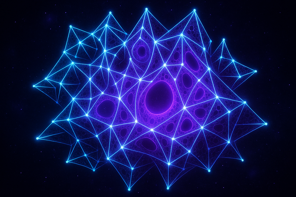
When the Blue Brain Project applied algebraic topology to its simulated cortical column, it found structures in up to 11 dimensions — cavities, cliques, and complexes that standard connectivity analysis had missed entirely. What those structures do is still an open question.
The Blue Brain Project: Topology of Neural Circuits
Series: Topological Data Analysis in Neuroscience | Part: 3 of 9
In 2017, a team of neuroscientists and mathematicians published a paper that should have broken the internet. They'd discovered that the rat cortex—not an abstract model, not a metaphor, but actual biological tissue—was computing in eleven-dimensional geometric space.
Not eleven neurons. Eleven topological dimensions.
Structures that have no physical analogue, that exist only in abstract mathematical spaces, were spontaneously assembling themselves from the coordinated firing of brain cells. Cavities. Voids. High-dimensional holes that form, persist, collapse, and reform as the network processes information.
This wasn't speculation. This was direct measurement. And it came from the most ambitious neuroscience project most people have never heard of: the Blue Brain Project—Henry Markram's attempt to simulate an entire piece of cortex in comprehensive molecular detail.
What they found when they turned it on and looked at it through topological eyes changed how we understand neural computation entirely.
What Blue Brain Actually Built
The Blue Brain Project started in 2005 with an audacious goal: reconstruct a cortical column from rat somatosensory cortex with such precision that you could run it as a simulation and watch real brain dynamics emerge.
Not a simplified model. Not a cartoon approximation. A digital reconstruction of biological tissue accurate down to individual neurons, complete with:
- The exact 3D positions of 31,000 neurons
- The precise morphology of each neuron's dendritic tree
- The specific locations of 8 million synapses
- The distribution of ion channels across each cell
- The synaptic dynamics—how neurotransmitters release, diffuse, bind
- The intrinsic electrical properties of each neuron type
They extracted this from actual rat brain slices, using electron microscopy, patch-clamp recording, immunohistochemistry, and every other technique that could measure microscopic detail. Then they assembled it into a computational model that ran on supercomputers—not to simplify the biology but to preserve it.
Why this matters: when you simulate brain tissue at this level of detail, you can't sneak in assumptions. The model doesn't "know" what it's supposed to do. It just follows physics. If patterns emerge, they emerge because that's what the biological architecture actually produces.
And when Markram's team stimulated their digital cortex with inputs mimicking sensory information, it started doing something nobody predicted.
It built geometry.
Enter the Topologists
Henry Markram had detailed neural simulations. Kathryn Hess had expertise in algebraic topology. Ran Levi had experience applying topological methods to complex systems. When they combined forces, the question was simple: What is the actual geometric structure of neural activity in this simulated tissue?
They applied persistent homology to the Blue Brain network. Not to the activity patterns (that came later), but first to the anatomical connectivity—the physical network of who connects to whom.
Standard neuroscience would represent this as a graph: neurons are nodes, synapses are edges, analyze with graph theory. Count degree distributions, measure clustering coefficients, find motifs.
TDA asked a different question: What topological structures exist in this network?
The answer was stunning.
The connectome—the wiring diagram of synaptic connections—contained directed simplicial complexes of remarkable sophistication. Not just edges (neuron A connects to neuron B). Not just triangles (A→B, B→C, C→A forming a loop). But tetrahedra, 5-simplices, 6-simplices, and all the way up to 11-dimensional structures.
These weren't sparse, random occurrences. They were organized. They were numerous. They were nested within each other in precise hierarchical patterns. The anatomical structure of the cortex had geometric depth that graph theory couldn't see.
And here's what made it even more remarkable: these geometric structures weren't static artifacts of the wiring diagram. They were functional. When the network was stimulated, these high-dimensional simplices became active—their constituent neurons fired in coordinated patterns that filled the geometric structures with activity.
The cavities came alive.
The Eleven Dimensions of Rat Cortex
Let's be precise about what they found.
When you stimulate the Blue Brain network with input, neurons fire in sequences. Not randomly—in structured cascades. As the activity propagates, it recruits neurons that form simplicial complexes. These complexes have dimension in the topological sense.
A 0-dimensional complex is a point (a single active neuron).
A 1-dimensional complex is an edge (two neurons that fired in sequence).
A 2-dimensional complex is a triangle (three neurons forming a directed loop of activation).
A 3-dimensional complex is a tetrahedron (four neurons in coordinated relationship).
And so on.
The Blue Brain researchers found that complex stimuli recruited directed cliques—groups of neurons all connecting to each other in specific patterns—up to dimension 11. Not occasionally. Systematically. Reproducibly.
These high-dimensional cliques had a property called cavities. A cavity is what you get when a shell of neurons fires coordinately but nothing in the center fires. It's a hole in activity space, bounded by active neurons. The cavity exists as an absence—a structured void.
And critically: the number and dimension of these cavities correlated with stimulus complexity.
Simple stimuli produced low-dimensional structures. Complex, information-rich stimuli produced high-dimensional cavities. The network was literally building geometric complexity in proportion to informational complexity.
This is not what anyone expected neural computation to look like.
Betti Numbers: Counting the Holes
To quantify this, they computed Betti numbers—the topological invariants that count holes in each dimension.
β₀ counts connected components (0-dimensional "holes"—i.e., separate pieces).
β₁ counts loops (1-dimensional holes).
β₂ counts voids (2-dimensional holes—like the hollow interior of a sphere).
β₃ counts 3-dimensional cavities.
...and so on.
For the Blue Brain network stimulated with complex inputs, they found non-zero Betti numbers up to dimension 7. That means the network was forming 7-dimensional topological features in its activity patterns. The anatomical structure supported even higher dimensions—up to 11.
To be clear: these dimensions don't correspond to physical space. You can't point to the 7th dimension in your brain. These are dimensions in the abstract space of possible firing patterns. The network state-space—the landscape of all possible configurations of which neurons are firing and which aren't—has geometric structure. And that structure has depth.
Different inputs carved different paths through this high-dimensional landscape, creating different topological features. The pattern of cavities encoded information about what the network was processing.
The topology was the computation.
Why Cavities Matter: Information and Topology
Here's the mechanistic insight: cavities aren't idle byproducts. They're computational primitives.
When a group of neurons forms a high-dimensional clique with a cavity at the center, that structure has specific informational properties:
1. Integration without centralization. The neurons around the cavity are all coordinated, but there's no central node. Information integrates across the group without flowing through a bottleneck.
2. Stability through redundancy. The cavity persists even if individual neurons fail. Remove one neuron from an 11-dimensional structure, and you still have a 10-dimensional structure. The information degrades gracefully.
3. Nonlinear amplification. Small changes in input can create or collapse entire cavities, producing nonlinear responses to stimulation. The geometry itself becomes a computational resource.
4. Temporal structure. Cavities form, persist for some duration, then collapse as the network state evolves. The lifespan of a cavity carries information—just like persistent homology tracks how long features last across scales.
This connects to something fundamental about how brains work. Neural computation isn't just about which neurons fire. It's about the pattern—the geometric structure of firing across populations. And that structure has properties you can't see by counting spikes or measuring pairwise correlations.
You need topology.
From Simulation to Biology
The obvious question: is this real, or is it a simulation artifact?
Fair concern. Computational models can produce spurious patterns. Maybe the high-dimensional cavities are bugs in the code, not features of biology.
The Blue Brain team addressed this rigorously. They compared their reconstruction to randomized networks with the same basic statistics—same number of neurons, same average connectivity, same degree distribution—but with connections rewired randomly.
Result: the random networks had drastically simpler topology. They formed low-dimensional structures, few cavities, minimal geometric depth. The specific wiring pattern of the biological cortex—not just its statistics—was necessary to produce the rich topological structure.
Moreover, when they compared different cortical regions and different developmental stages, they found systematic differences in topological structure. Visual cortex had different Betti numbers than motor cortex. Mature tissue had more complex topology than developing tissue.
The topology correlated with function.
And critically: subsequent work on actual biological recordings—not simulations—confirmed the findings. Multi-electrode arrays recording from real neural tissue show the same signatures. Calcium imaging capturing population activity in living brains shows topological features consistent with the Blue Brain predictions.
The cavities are real. The high-dimensional structures exist. The geometry is biology, not artifact.
What This Means for Neural Coding
Classical neuroscience thinks about neural coding in terms of rate codes (how fast neurons fire) or temporal codes (precisely when neurons fire). Both focus on individual neurons or small groups.
The Blue Brain discovery reveals a topological code: information encoded in the geometric structure of population activity.
This explains several mysteries:
Why population codes are so robust. If information lives in topology, it's preserved despite individual neuron variability. The geometry is the same even if specific neurons vary their firing rates.
Why the same neurons participate in different computations. A neuron can be part of different simplicial complexes at different times, contributing to different topological structures depending on context.
Why dimensionality reduction works. Techniques like PCA often reveal that high-dimensional neural data occupies low-dimensional manifolds. Those manifolds have topology. Reducing dimensionality preserves topology if you do it right.
Why damage can be so variable. Losing neurons destroys some simplices but not others. If critical geometric structures are preserved, function persists. If they're disrupted, even small damage causes large deficits.
The brain is computing with shapes in spaces we can't visualize. TDA lets us see those shapes anyway.
Implications for Coherence Geometry
Everything the Blue Brain Project found supports AToM's core framework:
1. Neural coherence is geometric. The integrated activity of neuronal populations creates persistent topological structure. High-dimensional cavities are literal instantiations of coherence—integrated patterns that maintain themselves across time.
2. Information is structure-preserving transformation. When the network processes input, it doesn't just change firing rates. It transforms the topology of its state space. Information flow is geometric flow. Computation is manifold evolution.
3. Complexity scales with topological richness. M = C/T predicts that meaning-making capacity scales with coherence. The Blue Brain data shows this explicitly: more complex stimuli produce higher-dimensional topological features. Meaning is encoded in geometric complexity.
4. Pathology is topological disruption. If neural computation depends on these geometric structures, then diseases that destroy connectivity should produce topological deficits. And indeed, neurodegenerative diseases progressively simplify the topology—cavities collapse, dimensions shrink, geometric richness degrades.
5. Scaling principles exist. The same mathematical tools that reveal topology in simulated cortex apply to whole-brain networks, to developmental trajectories, to cross-species comparisons. Topology is scale-invariant. The geometric principles operating at the level of cortical microcircuits operate at every level where coherence matters.
The Blue Brain Project proved something profound: coherence is not metaphor. It's measurable geometry.
This is Part 3 of the Topological Data Analysis in Neuroscience series, exploring how geometric methods reveal the hidden structure of mind.
Previous: Persistent Homology 101: Finding Features That Matter
Next: Topological Signatures of Consciousness: What Shape Is Awareness?
Further Reading
- Reimann, M. W., et al. (2017). "Cliques of neurons bound into cavities provide a missing link between structure and function." Frontiers in Computational Neuroscience, 11, 48.
- Markram, H., et al. (2015). "Reconstruction and simulation of neocortical microcircuitry." Cell, 163(2), 456-492.
- Hess, K., et al. (2017). "Algebraic topology methods in neuroscience." Notices of the American Mathematical Society, 64(10), 1107-1109.
- Sizemore, A. E., et al. (2018). "Cliques and cavities in the human connectome." Journal of Computational Neuroscience, 44(1), 115-145.
- Giusti, C., et al. (2015). "Clique topology reveals intrinsic geometric structure in neural correlations." Proceedings of the National Academy of Sciences, 112(44), 13455-13460.
Comments ()